Lower Leg Bone Diagram ~ įgalinti Sienas Priesiskumas Human Leg Bones Yenanchen Com. Your legs are two of your most impo. The bones of the leg and foot form part of the appendicular skeleton that supports the many muscles of the lower limbs. Health diagram bone skeleton leg knee science anchor chart human human body. Legs are used for standing, and all forms of. Sensory nerves are of course present throughout the lower extremities;
The myology of the lower limb is also particularly well represented in this atlas of anatomy, with multiple anatomical charts and diagrams: The quadriceps muscles straighten the knee. The lower leg contains two major long bones, the tibia and the fibula, which are both very strong skeletal structures. Lower jaw (mandible) collar bone. The tibia (also called the shinbone) is located near the midline of the leg.
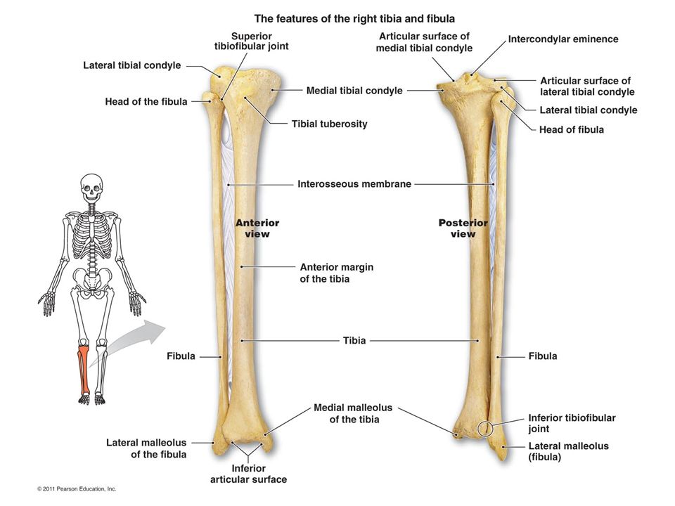
8 4 Bones Of The Lower Limb Anatomy Physiology from open.oregonstate.education Our goal is that these leg anatomy worksheets pictures gallery can be a direction for you, bring you more references and also make you have a great day. The largest and most medial leg bone, forming both the knee and ankle joints. In this video i talked about lower limb anatomy which includes bones such as pubis, tibia, patella, fibula and laos joints such as knee joint and ankle joint. As pain is the body's way of telling you something is wrong and it needs to heal, it is wise to pay attention to this pain rather than fight through it. The lower leg contains two major long bones, the tibia and the fibula, which are both very strong skeletal structures. The head of the fibula. The hip joint gives the leg an incredible range of motion while still providing support to the body's weight. At the distal end of the femur, two rounded condyles meet the tibia and fibula bones of the lower leg to form the knee joint.
Diagram and names of leg bones, diagram of foot and leg bones, diagram of leg bones, diagram of lower leg bones, diagram of the bones in your leg, bone, diagram and.
Health diagram bone skeleton leg knee science anchor chart human human body. This large tendon from the powerful thigh muscles (quadriceps) wraps round the patella and is attached to the top of the lower leg bone (tibia). The lower leg extends from the knee to the ankle. Beside that, we also come with more related ideas as follows free printable human anatomy coloring pages, lower leg muscle diagram blank and lower limb bones unlabeled. The nerves of the leg and foot serve to propel the body through the actions of the legs, feet, and toes while maintaining balance, both while the body is moving and when it is at rest. See more ideas about muscle anatomy, human anatomy and physiology, body anatomy. This area is commonly referred to as the calf. He leg's main function in the human is for locomotion and support of the rest of the body. Most leg pain results from wear and tear, overuse, or injuries in joints or bones or in muscles, ligaments, tendons or other soft tissues. The largest and most medial leg bone, forming both the knee and ankle joints. The thigh bone, or femur, is the large upper leg bone that connects the lower leg bones (knee joint) to the pelvic bone (hip joint). The lower leg contains two major long bones, the tibia and the fibula, which are both very strong skeletal structures. The foot bones shown in this diagram are the talus, navicular, cuneiform, cuboid, metatarsals and calcaneus.
The lower leg consists of muscles, bones, tendons, ligaments, joints, blood vessels, and nerves. Legs are used for standing, and all forms of. Damage or irritation to any of these structures can lead to lower leg pain. Sensory nerves are of course present throughout the lower extremities; The largest and most medial leg bone, forming both the knee and ankle joints.
Foot Ankle Unit Skeletal Anatomy Lower Leg Bones Tibia Shin Bone Weight Bearing Bone In The Lower Leg Medial Malleolus Distal End Of Tibia That Ppt Download from images.slideplayer.com This keeps the bones together, giving a high ankle sprain time to heal. The medial side of the tibia is located immediately under the skin, allowing it to be easily palpated down the entire length of the medial leg. Bone diagram forehead (frontal bone) nose bones (nasals) cheek bone (zygoma) upper jaw (maxilla) lower jaw (mandible) breast bone (sternum) upper arm bone (humerus) lower arm bone (ulna) thigh bone (femur) collar bone (clavicle) toe bones (phalanges) ankle bones (tarsals) kneecap (patella) shin bone (tibia) calf bone (fibula) foot bones The head of the femur forms the ball and socket hip joint with the acetabulum of the hip bone. See more ideas about muscle anatomy, human anatomy and physiology, body anatomy. The lower leg contains two major long bones, the tibia and the fibula, which are both very strong skeletal structures. The major bones of the leg are the femur (thigh bone), tibia (shin bone), and adjacent fibula, and these are all long bones.the patella (kneecap) is the sesamoid bone in front of the knee.most of the leg skeleton has bony. Learn vocabulary, terms, and more with flashcards, games, and other study tools.
The largest and most medial leg bone, forming both the knee and ankle joints.
Health diagram bone skeleton leg knee science anchor chart human human body. The foot bones shown in this diagram are the talus, navicular, cuneiform, cuboid, metatarsals and calcaneus. Sensory nerves are of course present throughout the lower extremities; Your legs are two of your most impo. Legs are used for standing, and all forms of. However, with the exception of the bottom of the foot, they play a lesser role here than. Most leg pain results from wear and tear, overuse, or injuries in joints or bones or in muscles, ligaments, tendons or other soft tissues. Bone diagram forehead (frontal bone) nose bones (nasals) cheek bone (zygoma) upper jaw (maxilla) lower jaw (mandible) breast bone (sternum) upper arm bone (humerus) lower arm bone (ulna) thigh bone (femur) collar bone (clavicle) toe bones (phalanges) ankle bones (tarsals) kneecap (patella) shin bone (tibia) calf bone (fibula) foot bones The medial side of the tibia is located immediately under the skin, allowing it to be easily palpated down the entire length of the medial leg. The patella is the kneecap bone. The head of the femur forms the ball and socket hip joint with the acetabulum of the hip bone. Diagram and names of leg bones, diagram of foot and leg bones, diagram of leg bones, diagram of lower leg bones, diagram of the bones in your leg, bone, diagram and. In this video i talked about lower limb anatomy which includes bones such as pubis, tibia, patella, fibula and laos joints such as knee joint and ankle joint.
Our goal is that these leg anatomy worksheets pictures gallery can be a direction for you, bring you more references and also make you have a great day. Start studying pelvis, leg bones, leg bones. This keeps the bones together, giving a high ankle sprain time to heal. We'll break down the anatomy and function of the upper leg, knee, lower leg, ankle, and foot. The head of the femur forms the ball and socket hip joint with the acetabulum of the hip bone.
Bones Of The Upper Limb Anatomy Physiology from pressbooks-dev.oer.hawaii.edu The back of the patella is covered with smooth cartilage. The nerves of the leg and foot serve to propel the body through the actions of the legs, feet, and toes while maintaining balance, both while the body is moving and when it is at rest. The first diagram summarizes the different muscular compartments (fascial compartments) of the thigh and leg, and the different fascias (crural fascia, intermuscular septum, interosseous membrane, adductor canal, fascia lata) Lower jaw (mandible) collar bone. Learn vocabulary, terms, and more with flashcards, games, and other study tools. As pain is the body's way of telling you something is wrong and it needs to heal, it is wise to pay attention to this pain rather than fight through it. The medial side of the tibia is located immediately under the skin, allowing it to be easily palpated down the entire length of the medial leg. See more ideas about muscle anatomy, human anatomy and physiology, body anatomy.
The largest and most medial leg bone, forming both the knee and ankle joints.
The thigh bone, or femur, is the large upper leg bone that connects the lower leg bones (knee joint) to the pelvic bone (hip joint). Bone diagram forehead (frontal bone) nose bones (nasals) cheek bone (zygoma) upper jaw (maxilla) lower jaw (mandible) breast bone (sternum) upper arm bone (humerus) lower arm bone (ulna) thigh bone (femur) collar bone (clavicle) toe bones (phalanges) ankle bones (tarsals) kneecap (patella) shin bone (tibia) calf bone (fibula) foot bones The foot bones shown in this diagram are the talus, navicular, cuneiform, cuboid, metatarsals and calcaneus. The nerves of the leg and foot serve to propel the body through the actions of the legs, feet, and toes while maintaining balance, both while the body is moving and when it is at rest. The patella is the kneecap bone. The tibia (also called the shinbone) is located near the midline of the leg. The lower leg is comprised of two bones, the tibia and the smaller fibula. The major bones of the leg are the femur (thigh bone), tibia (shin bone), and adjacent fibula, and these are all long bones.the patella (kneecap) is the sesamoid bone in front of the knee.most of the leg skeleton has bony. The quadriceps muscles straighten the knee. At the distal end of the femur, two rounded condyles meet the tibia and fibula bones of the lower leg to form the knee joint. Your legs are two of your most impo. The fibula or calf bone is a leg bone on the lateral side of the tibia, to which it is connected above and below.it is the smaller of the two bones and, in proportion to its length, the slenderest of all the long bones. The head of the femur forms the ball and socket hip joint with the acetabulum of the hip bone.
Ebraheim's educational animated video describes the muscle and nerve anatomy of the lower legthere are fourteen muscles within the lower leg leg bone diagram. Diagram and names of leg bones, diagram of foot and leg bones, diagram of leg bones, diagram of lower leg bones, diagram of the bones in your leg, bone, diagram and.
Share :
Post a Comment
for "Lower Leg Bone Diagram ~ įgalinti Sienas Priesiskumas Human Leg Bones Yenanchen Com"


Post a Comment for "Lower Leg Bone Diagram ~ įgalinti Sienas Priesiskumas Human Leg Bones Yenanchen Com"